Electrocardiography (ECG or EKG) is a process that measures the electrical activity of the heart through sensors called electrodes. Here’s an overview of how it works:
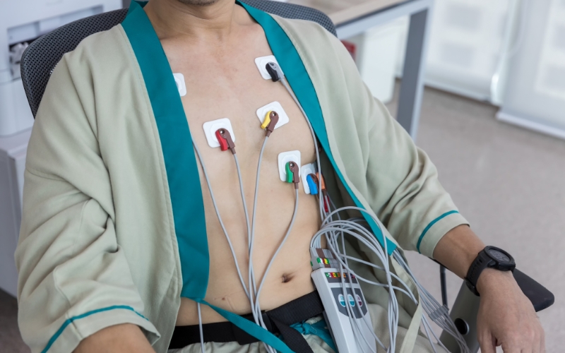
Electrode Placement: Several electrodes are placed on the skin, usually on the chest, arms, and legs. These electrodes detect electrical signals generated by the heart during its beating cycle.
Signal Detection: The heart produces electrical impulses during each heartbeat. These impulses travel through the electrodes to the ECG machine. Each electrode detects changes in electrical potential from different angles.
Data Transmission: The collected signals are transmitted to the ECG machine, where they are processed and displayed as a waveform.
Waveform Analysis: The ECG waveform consists of different parts (P wave, QRS complex, T wave) that represent various phases of the cardiac cycle, such as atrial depolarization, ventricular depolarization, and repolarization.
Interpretation: The recorded waveforms are analyzed by a physician to detect abnormalities like arrhythmias, ischemia, or heart damage.
ECG is non-invasive, quick, and a critical tool for diagnosing heart conditions. It can identify problems like heart attacks, irregular rhythms, and other heart-related issues, guiding effective treatment.