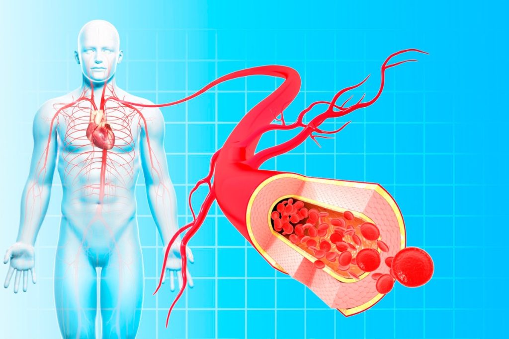
Angiography is a medical imaging technique used to visualize the inside of blood vessels and organs of the body. It is primarily used to diagnose and evaluate conditions related to the vascular system, including arteries, veins, and capillaries. Here’s an overview of how angiography works and its key applications:
How angiography typically works
Contrast Agent Injection:
Process: A contrast dye, which is a special fluid that enhances the visibility of blood vessels, is injected into the bloodstream through a catheter. The catheter is typically inserted into a large artery, often in the groin or arm, and carefully guided to the area of interest.
Purpose: The contrast dye highlights the blood vessels and allows them to be clearly visible on the imaging system.
Imaging:
Procedure: Once the contrast dye is injected, a series of X-ray images or other types of imaging (such as CT or MRI) are taken. These images capture detailed views of the blood vessels and any potential abnormalities.
Technique: Depending on the type of angiography, different imaging techniques may be used. Conventional angiography uses X-rays, while CT angiography uses computed tomography, and MRI angiography employs magnetic resonance imaging.
Analysis:
Evaluation: The resulting images are analyzed by a radiologist or cardiologist to assess the condition of the blood vessels. They look for issues such as blockages, narrowing, aneurysms, or malformations.
Diagnosis: Based on the findings, a diagnosis can be made, and appropriate treatment plans can be developed.
Uses of Angiography
Angiography is a versatile imaging technique with several critical uses in medical diagnostics. Primarily, it is employed for cardiac evaluation to assess the coronary arteries, helping to diagnose coronary artery disease (CAD) and guide treatments like angioplasty or stent placement. It is also vital in stroke diagnosis, where it visualizes blood vessels in the brain to identify causes such as aneurysms, arteriovenous malformations (AVMs), or clots, which is essential for timely and effective treatment. Additionally, angiography is used to evaluate peripheral artery disease (PAD) by examining blood vessels in the limbs, aiding in the management of arterial blockages and improving blood flow. In cases of pulmonary embolism, angiography helps detect blockages in the lung arteries, facilitating prompt intervention. Furthermore, it plays a crucial role in pre-surgical planning, providing detailed vascular images to assist surgeons in planning and executing procedures with precision. Lastly, it helps in detecting and managing vascular malformations and aneurysms, allowing for accurate diagnosis and effective treatment. Overall, angiography provides detailed insights into the vascular system, guiding treatment decisions and enhancing patient care.
Dr. Alok Shah, a renowned cardiologist, excels in diagnosing and treating heart conditions with expertise and compassionate care.